The inauguration ceremony was attended by university authorities, academics and students. ‘The fact that we are inaugurating this FONDEQUIP today is due to the existence of BIOREN UFRO, the installed capacities within the University, and most importantly, this technology will be of use to many researchers’, stated the Rector of UFRO, Dr Juan Manuel Fierro.
The acquisition of this microscope forms part of a broader scientific development strategy. The BIOREN-UFRO TEM is the result of a collaborative research effort supported by seven partner institutions: the Universidad de Aysén, the Universidad Católica de la Santísima Concepción, the Universidad Católica de Temuco, the Universidad Santo Tomás, the Centro de Estudios Científicos (CECs)-Valdivia, and the INIA Carillanca Regional Research Centre. ‘To address critical global challenges, we must understand and manipulate matter at the atomic level, which requires the use of advanced technologies such as the Talos L120C G2 Transmission Electron Microscope’, explained Dr Marcela Calabi.
Among the researchers and collaborators involved are Dr Jaime Mejías (Instituto de Investigaciones Agropecuarias, INIA Carillanca); Dr Felipe Barros (Faculty of Medicine, Universidad Santo Tomás, and National Director of Research at CECs); Dr Carla Basualto (Department of Health Sciences, Universidad de Aysén); Dr Fabiola Valdebenito (Faculty of Sciences, Universidad Católica de la Santísima Concepción); and Dr Jacobo Hernández (Head of the Department of Mathematical and Physical Sciences, Universidad Católica de Temuco).
‘Collaboration is essential to advance complex issues, as not all the answers, specific knowledge or experience within research lines are always available. This is why collaboration and the promotion of strategic partnerships are so important. This project involves not only BIOREN but also outstanding scientists from across the country’, stated Dr María de la Luz Mora, Director of the Scientific and Technological Bioresource Nucleus, BIOREN UFRO.
Nanometric-scale research
Unlike an optical microscope, which uses light, a TEM uses a beam of electrons that passes through samples that must be ultrathin. This allows for the visualisation of subcellular structures, such as specific organelles, as well as the characterisation of soft materials including polymers, nanocomposites and nanoparticles.
In terms of specific applications, this equipment will strengthen research related to the development of smart fertilisers, sustained-release nanocomposites and environmentally friendly nanopesticides; the tracking of morphological changes in plant chloroplasts under conditions of extreme water stress; ultrastructural analysis in dentistry, reproduction and biomedicine; and the characterisation of polymers and new materials for engineering.
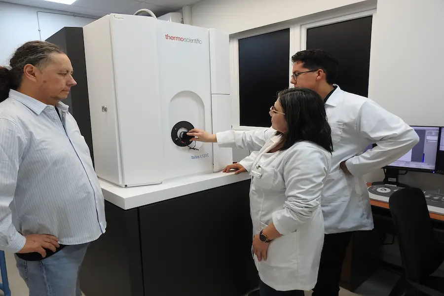
The equipment is integrated into the Microscopy and Flow Cytometry Unit, led by Dr Karina Godoy. Its operation is overseen by electronic civil engineer José Soto and dentist Alejandra Valle. This diversity of professional backgrounds ensures a comprehensive interpretation of data and expert support for researchers.
Written by BIOREN, Universidad de La Frontera